Description
We develop a simplified PS-OCT device (called polarization-diversity OCT) tailored for clinical imaging of choroidal tumor, which generate additional contrast of pigment-containing structures in the eye.
Optical Imaging System
Schematic of the 400kHz polarization-diversity optical coherence tomography (PD-OCT) system
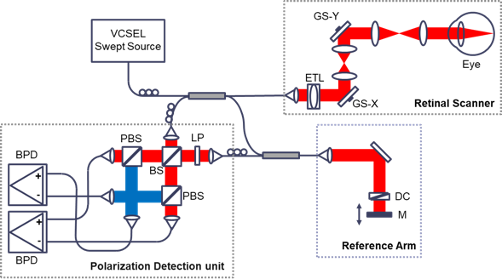
M: mirror; PC: polarization controller; DC: dispersion
compensation block; LP: linear polarizer; BS: beam splitter; PBS: polarization beam splitter; L1-
L4: lens; ETL: electrical tunable lens; GS-X and –Y: x-axis and y-axis galvanometer scanner; Vand H-BPD: balanced photodetector for horizontally and vertically polarized signals.
| |
Parameter |
| Wavelenght (nm) |
1060 |
| Bandwith (nm) |
100 |
| A-scan rate (Hz) |
400,000 |
| Power at cornea (mW) |
1.5 |
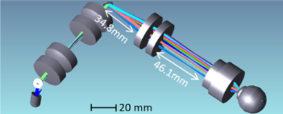
Ultrawidefield retinal scanner
In collaboration with Prof. Yifan Jian, we designed a ultrawide retinal scanner.
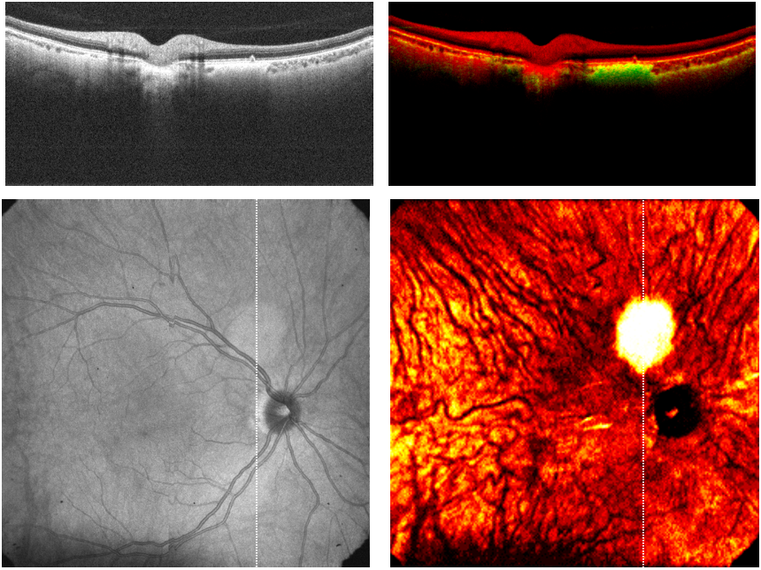
Results
OCT intensity (left) and depolarization (right) image
Healthy subject
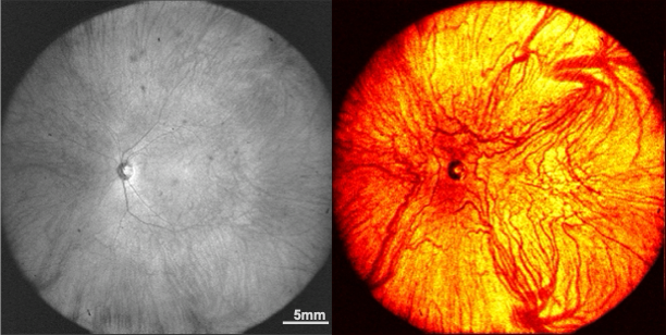

Subject with melanotic choroidal tumor
References
Y. Miao, H. Jung, D. Hsu, J. Song, S. Ni, D.. Ma, Y. Jian, S. Makita, Y. Yasuno, M.V. Sarunic, K.A.J. Stephen-
son, K. Paton, Z. Mammo, M.J. Ju. (2023). Polarization-Diversity Optical Coherence Tomography Assessment of Choroidal Nevi, Investigative Ophthalmology & Visual Science. 64(14):6. doi: 10.1167/iovs.64.14.6.